International Medical Conference
Endometriosis 2024:
Elevating Sampson’s Century Legacy via
Deep Dive with AI
For the benefit of Endometriosis Foundation of America (EndoFound)
May 2-3, 2024 - JAY CENTER (Paris Room) - NYC
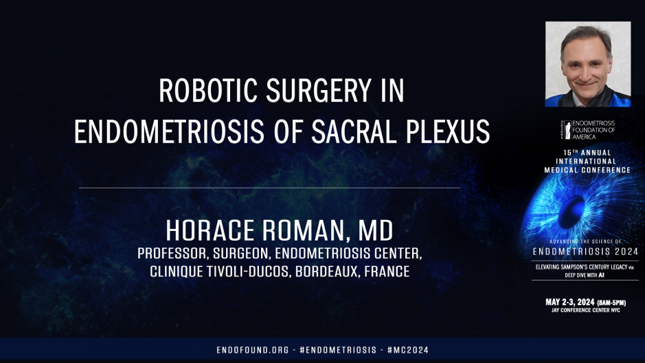
Ladies and gentlemen, dear Colleague, dear friends, it is my absolute pleasure to be with you virtually during this prestigious meeting and very grateful to my friends Tam and Dan Martin for this kind invitation. Unfortunately, due to family problems, I cannot be with you in person. However, I will share with you my thoughts about the place of the robotic surgery in the management of endometriosis of sacral plexus. I have some conflicts of interest because I'm routinely involved in training, meetings and workshops. However, this is without impact on my own thoughts. The sacral plexus is located into the parum together with the ureter and the internal iliac vessels, it is represented by large nerves giving birth to thin nerves. Large nerves measure from five kilometer to one centimeter. They are easy to identify and they are somatic. They should not be injured because it leads to anesthesia and motor pal thinners or vegetative or autonomic.
They control the function of rectum, bladder, and vagina. They are much more difficult to identify and to preserve when they are involved by deep endometriosis nodules. The technique of nerve sparing specifically refer to this thin nerves in order to avoid pelvic organ dysfunction. There are two major types of nodules infiltrating the parum, the most frequent or close to the midline. They involve in majority the last three sacral roots, but also the rectum on the vagina, and they are responsible for vegetative troubles such as the bladder voiding dysfunction. This nodules always involve the inferior hypogastric plexus, which explain bladder and rectal dysfunction. Conversely, other nodules are located more laterally on contact with the lateral pelvic wall and they involve the lumbosacral trunk and the sciatic nerve. Thus, they are responsible for somatic troubles such as somatic pain or walking troubles. While the bowel and bladder dysfunction may be absent, indeed, death nodules may be lateral from the inferior gastric plexus and may not involve autonomic nerves.
Now, clinical nerve dysfunction may have two origins. The endometriosis itself may involve and destroy the nerves, but also the surgery may affect the nerves. In most of cases, postoperative dysfunction is reversible because it is due to the neuropraxia following the dissection or the al spread during the surgery. While in some cases the nerves may be destroyed or uced by the surgeon in this case, the troubles or definitive. Here you have baseline symptoms of 100 patients. We managed at Ifdo Bodo during four year and a half. This patient out of 10 had bladder void dysfunction before the surgery, as well as severe constipation and various degrees of sciatica. They underwent complex surgical procedure, which required the dissection of scra roots in all the cases and that of the satic nerve. In one woman out of five, two women out of three required a colorectal resection of disc excision.
One woman out of three had a ureter involvement and one woman out of seven had urinary tract sutures. Definitively woman presenting with endometriosis involving sacral preuss require in a majority of cases a multidisciplinary surgical approach. We published two 10 step video articles where we detailed the surgical approach on sacral roots and satic nerve in women with large nodules involving the medial or lateral parum. This articles are in open access, so I would invite you to have a look at them as rga. The preservation of thin autonomic nerves by nerves sparing it is best described by the Negra technique, which was introduced by the team of macaroni and luli, and includes five standardized steps with the successive dissection of superior hippo gastric plexus, hippo gastric nerve, inferior hippo, gastric plexus, and splenic nerves in order to preserve rectal and bladder function. Since June, 2021, we have completely stopped to perform the excision of deep endometriosis of the parameter in conventional laparoscopy, and we have moved to the robotic surgery which provides some major advantages such as the use of sharp and miniaturized instrument, which allows a more precise dissection.
The instruments have seven degree of freedom, which allows a tangential approach on nerves and the full access of small deep spaces. The death of the view is improved and stable, which allows, in my opinion, a better identification of thin nervous fibers. Here you have an example of excision of a deep endometriosis nodule involving the sacral roots, particularly the S3 and S four. We follow the 10 step procedures described in our video article published in fertility and sterility. Advantages of robotic assistance are obvious during the challenging step of the control of internal iliac artery and vein. Some of their major branches should be clipped and sectioned in order to reveal the sacral roots, which are located behind the vessels and in the front of peripheral muscle. Thanks to the mobility of instruments, the clipse are placed perpendicular on the vessels while the dissection of sachar root is done tangentially, which reduces in my opinion, the risk of injury of superficial fibers. The view is stable and there is no conflict between the endoscope and the straight instruments as we may have in laparoscopy.
This allows a precise dissection of tissues and increases, in my opinion, the probability of preservation of thin fibers, which are not involved by the disease. This step usually takes 30 to 60 minutes. Let's see now the excision using the robotic assistance of type two nodules located on contact with the lateral pelvic wall and infiltrating the lumbosacral trunk and the satic nerve. The dissection is carried out medially from the external iliac vessels with identification of the nerve, which may be involved, which is located one or two centimeter proximally from the lumbosacral trunk. The articulation of robotic instruments allows a comfortable dissection in a very small space and progressive identification of lumbosacral trunk and sacral roots have to control several vessels such as the umbilical artery, but also observatory vessels in order to have a full access on the posterior limit of the nodule. Then the dissection is done progressively tangentially to sacral roots and to lobos trunk until the grid satic notch to employ a low setting of the monopolar current, which avoids the contraction of muscles of the leg each time the current is used proximal to the nerve.
The origin of autonomic splenic nerves is usually medial from the limit of the excision, which explains why the risk of bladder and rectal dysfunction is lower than that observed after the excision of type one parametal nodules. The penal nerve is routinely released during this procedure, which usually takes two to three hours. Such complex surgical procedures are logically related with some postoperative complications, some complications or related to the manipulation of nerves themselves, such as the bladder dysfunction, which may require a self catheterization for several weeks and months in one patient out of five other complications or related to additional procedures performed on the low and mid rectum such as rectal fistula, which rate may average eight to 10% a second surgery due to early postoperative complications may be performed in one patient out of five. Despite the risk of complications, the surgery is worthwhile because we can observe a rapid improvement of sciatica and pudendal pain.
Almost all patients postoperative lower limb motor weakness is very rare and usually progressively improves thanks to the physiotherapy. Patients satisfaction rate is very high even in those experiencing postoperative complications. In conclusion, we can affirm that deep nodules of parametria always involve some nerves. Somatic or autonomic, their excision is now well standardized and the robotic assistance is, in my opinion, most useful. The negro technique allows the preservation of autonomic nerves when they are not involved by the fibrile nodules, while our 10 step procedures to avoid intraoperative vessel or nerve injuries to enable a complete excision of deep endometriosis, s nodules, medial S lateral, and to allow training and exchanges between surgeons. Once again, I'm very grateful for this kind invitation. I'm proud to be virtually with you, and I am so sorry that due family problems I could not be with you in person. I wish you an excellent meeting and I am available for any questions.


